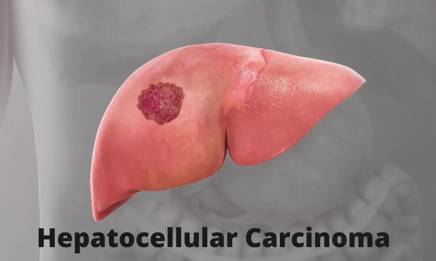
South Korea: A newly developed MRI-based scoring system may help identify patients at higher risk of early recurrence after surgical removal of small hepatocellular carcinoma (HCC). The findings, published in Radiology, suggest that the MRI-based Early Recurrence Individualized Score (MERIS) can more accurately predict early tumor recurrence following resection of solitary HCC lesions measuring 5 cm or smaller.The research was led by Eun Sun Choi from the Department of Radiology and Research Institute of Radiology at the University of Ulsan College of Medicine and Asan Medical Center in Seoul, Republic of Korea.Surgical resection remains the primary treatment for early-stage hepatocellular carcinoma. However, recurrence remains a major concern, occurring in nearly half of patients within two years of surgery and in up to 70% within five years. Because of this high recurrence rate, identifying individuals at greater risk of early relapse is important for guiding surgical planning, selecting candidates for transplantation, and considering additional perioperative therapies.Although several imaging-based models have been proposed to estimate recurrence risk, many were developed using mixed patient populations that included individuals with large or multiple tumors. As a result, their predictive value may be limited for patients with small solitary HCC, who may have different recurrence risk profiles.To address this gap, the investigators developed and validated MERIS, a predictive scoring system derived from MRI findings and clinical parameters. The study included 325 patients with treatment-naïve solitary HCC measuring 5 cm or less who underwent curative surgical resection and gadoxetic acid–enhanced liver MRI. Among them, 204 patients were included in the training dataset used to develop the model, while 121 patients formed an external validation cohort. The following were the key findings:Multivariable Cox regression analysis identified four factors associated with early recurrence within two years after surgery.These factors included elevated aspartate aminotransferase levels, larger tumor size, a nonsmooth tumor margin on imaging, and peritumoral hepatobiliary phase hypointensity on MRI.These variables were integrated into the MRI-based Early Recurrence Individualized Score (MERIS) model.The MERIS model demonstrated strong predictive performance, with a Harrell’s concordance index (c-index) of 0.75 in both the training and external validation sets.A cutoff score of five points effectively stratified patients into low- and high-risk recurrence groups.In the training cohort, the two-year recurrence-free survival rate was 91.4% in the low-risk group and 69.5% in the high-risk group.In the external validation cohort, the two-year recurrence-free survival rates were 87.4% in the low-risk group and 59.3% in the high-risk group.The MERIS model outperformed pathology-based prognostic models and several existing prediction systems in the external validation analysis.These findings highlight the potential of MRI-based risk stratification for identifying patients who may benefit from closer monitoring or additional therapeutic strategies.The authors noted that by providing more accurate preoperative assessment of recurrence risk, MERIS could assist clinicians in optimizing surgical planning, transplant decision-making, and perioperative treatment strategies. They emphasized that further prospective studies in broader patient populations will be needed to confirm the model’s generalizability and clinical utility.Reference: https://doi.org/10.1148/radiol.252611