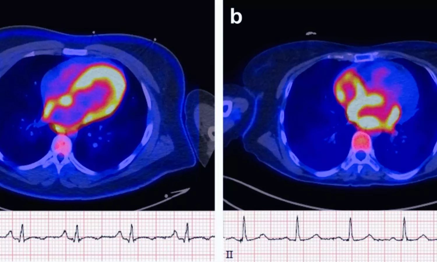
USA: Positron emission tomography (PET) imaging may help identify patients at higher risk of developing atrial fibrillation (AF) by detecting increased metabolic activity in the atrial walls, a new study published on January 20 in JACC: Advances has found. The findings suggest that atrial uptake of the F-18 fluorodeoxyglucose (FDG) radiotracer is associated with a significantly higher likelihood of incident AF over long-term follow-up.The study was led by Romanos Haykal, MD, from the Division of Cardiology, Department of Medicine, University of Washington, Seattle, and colleagues. AF is a common arrhythmia linked to an increased risk of stroke, heart failure, and mortality. Emerging evidence indicates that abnormal cardiac metabolism—particularly a shift toward increased glucose utilization—may contribute to the initiation and progression of AF. Given its ability to visualize glucose metabolism, FDG-PET imaging was hypothesized to provide insights into early metabolic changes preceding AF onset.The investigators analyzed data from 207 adults who underwent FDG-PET imaging at the University of Washington between 2017 and 2024, most commonly for evaluation of suspected cardiac sarcoidosis. Patients with prior AF, inadequate follow-up, or suboptimal image quality were excluded. Participants were followed for a median of 3.35 years to assess the development of new-onset AF.PET images were evaluated using both visual and semiquantitative methods. Atrial FDG uptake was assessed based on intensity relative to background activity, while standardized uptake values and target-to-background ratios were calculated in areas of highest uptake. The researchers reported the following findings:During follow-up, 50 patients (24.2%) developed new-onset atrial fibrillation.Patients who developed AF were older and had a higher prevalence of hypertension, coronary artery disease, and cardiac sarcoidosis compared with those who remained AF-free.Atrial FDG uptake was significantly more frequent in patients who developed AF than in those who did not.FDG uptake was observed more often in the atria than in the ventricles.Uptake was predominantly localized to the right atrium rather than the left atrium or both atria.Kaplan–Meier analysis showed an early and sustained separation in AF incidence between patients with and without atrial FDG uptake.Multivariable Cox regression confirmed atrial FDG uptake as a strong independent predictor of AF, associated with more than a threefold increase in risk after adjustment for age and cardiovascular comorbidities.The authors highlighted two key observations: a strong association between atrial FDG uptake and subsequent AF, and a predominance of right atrial metabolic activity. This pattern aligns with prior studies suggesting that the right atrium may be involved earlier in the atrial remodeling process. Alternatively, reduced metabolic activity in the left atrium due to fibrosis may contribute to the apparent right-sided dominance.Although the study population was enriched for patients undergoing evaluation for cardiac sarcoidosis, and asymptomatic AF episodes may have been missed, the findings highlight the potential role of PET imaging in AF risk stratification. Overall, the study suggests that atrial metabolic abnormalities detected by FDG-PET may serve as an early marker of AF susceptibility, opening avenues for improved prediction and prevention strategies.Reference:Haykal, R, Kassar, A, Chamoun, N. et al. Atrial 18FDG Uptake Predicts Incident Atrial Fibrillation in Patients Undergoing Cardiac PET Imaging. JACC Adv. 2026 Feb, 5 (2). https://doi.org/10.1016/j.jacadv.2025.102569
Atrial FDG Uptake on Cardiac PET Signals Higher Risk of Future Atrial Fibrillation: Study