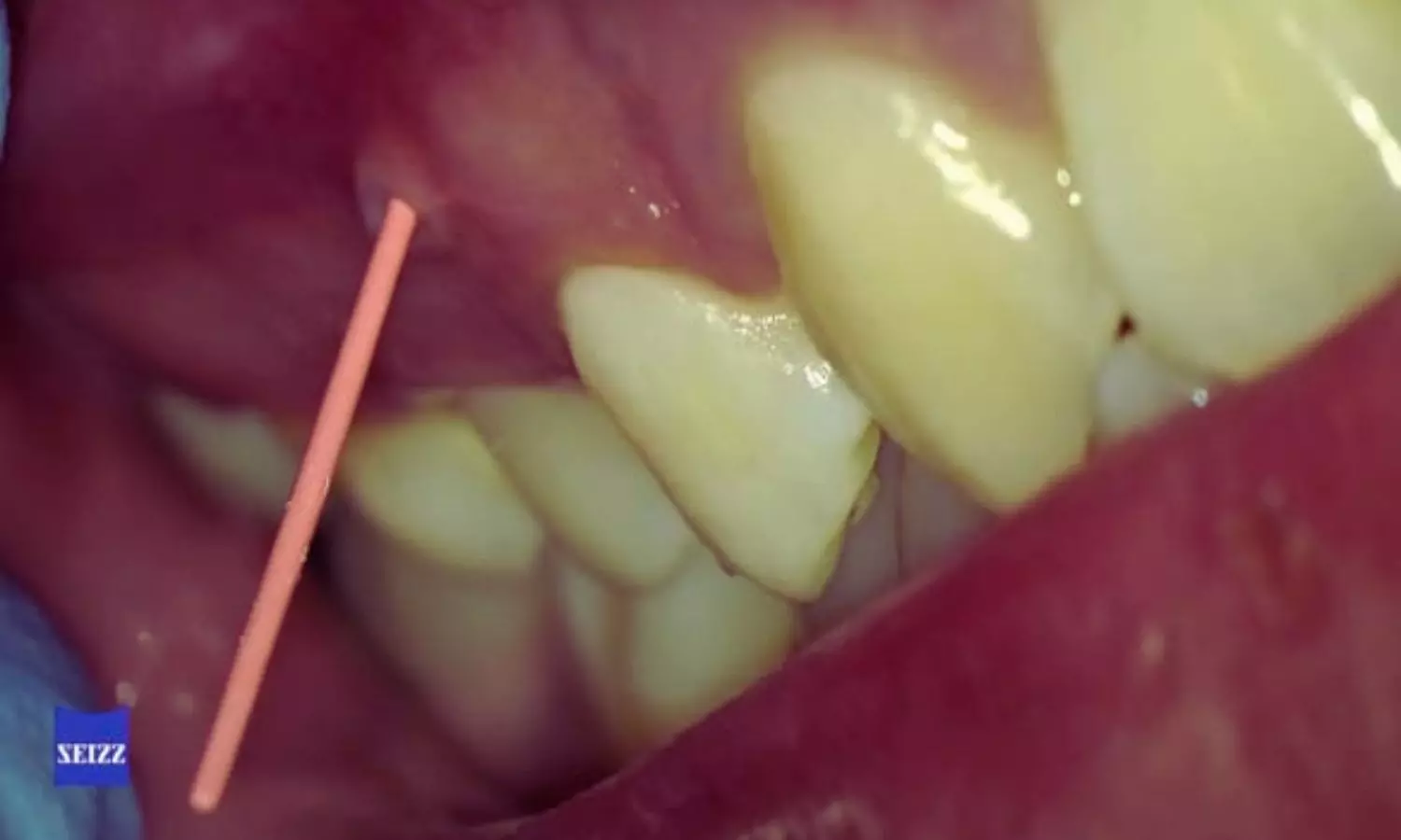
Brazil: The combination of periapical radiography (PR) and cone-beam computed tomography (CBCT) significantly improves diagnostic accuracy for both endodontists and periodontists. This integrated approach provides more reliable detection and assessment of complex cases compared to PR alone, supporting better clinical decision-making and interdisciplinary collaboration.A new study published in the Brazilian Dental Journal highlights the added diagnostic value of combining conventional periapical radiographs with cone-beam computed tomography when evaluating complex endodontic and periodontal lesions. The research, led by Maria Caroline Rios Piecha from the Graduate Program in Dentistry at the Universidade Federal de Pelotas, Brazil, examined how access to CBCT imaging influences diagnostic performance among dental specialists.Accurate diagnosis is critical in cases where pulp and periodontal tissues are both involved, as clinical and radiographic features often overlap. Traditional periapical radiography remains a first-line imaging modality in dental practice due to its accessibility and low radiation dose. However, its two-dimensional nature can limit visualization of intricate anatomical details, potentially leading to misinterpretation of complex lesions. CBCT, by contrast, provides three-dimensional imaging that can reveal structures and pathologies not clearly visible on standard radiographs.In the cross-sectional study, the investigators recruited 60 dental specialists—30 endodontists and 30 periodontists—who evaluated ten carefully selected clinical cases. These cases represented a range of challenging conditions, including vertical root fractures or fissures, endodontic perforations, persistent apical periodontitis, combined endo-periodontal lesions, and localized periodontitis. Each participant assessed the cases at two separate stages.Initially, the specialists reviewed clinical information alongside periapical radiographs and recorded their diagnostic hypotheses using a structured questionnaire. In the second stage, CBCT images of the same cases were added, allowing participants to reassess the findings and revise their diagnoses. For each case, the correct diagnosis—established by the dentist responsible for patient management—was included among the response options. Diagnostic accuracy before and after CBCT incorporation was statistically analyzed using the McNemar test. The study revealed the following findings:Adding CBCT to periapical radiography markedly improved diagnostic accuracy, increasing correct diagnoses from 42.2% with PR alone to 67.2% with the combined approach.When CBCT was available, both endodontists and periodontists achieved a diagnostic accuracy of 58.8%.The PR+CBCT combination was especially effective in identifying endo-periodontal lesions, including cases with and without associated root damage, compared with periapical radiography alone.The authors noted that, to their knowledge, this is the first study to assess the impact of PR combined with CBCT on diagnosing lesions involving both pulp and periodontal tissues, with participation from specialists in both disciplines. Earlier research largely focused on endodontists, underscoring the novelty and interdisciplinary relevance of the current findings.Within the study’s limitations, the authors concluded that integrating CBCT with periapical radiography enhances diagnostic accuracy in complex cases affecting pulp and periodontal tissues. The findings emphasize the importance of collaboration between endodontists and periodontists and support the use of combined imaging strategies to achieve more accurate diagnoses, clearer prognoses, and better-informed treatment planning.Reference: https://doi.org/10.1590/0103-644020256704